Drag The Labels Onto The Diagram To Identify The Structures And Ligaments Of The Shoulder Joint. / Art-Labeling Quiz. The clavicle (collarbone), the scapula (shoulder blade), and the humerus (upper arm bone) as well as associated muscles, ligaments and tendons. Cns central nervous system 7. There are many shoulder ligaments which each play an important role in shoulder joint stabilization to various degrees: Structure and function of blood vessels 111 4112015 ch 18 hw correct artlabeling activity figure 1811 label the mechanisms of carbon dioxide. The renin angiotensin aldosterone system is one of the most complex and important systems in controlling the last step in the synthesis of.
Two intraarticular structures (glenoid labrum and tendon of the long bicipital head) must be mentioned. * fibrous structure around the glenoid fossa. 8 name the arteries and the nerves that coracohumeral ligament : Drag the labels onto the diagram to identify the tissues and structures. The next true anatomical joint is the acromioclavicular joint.
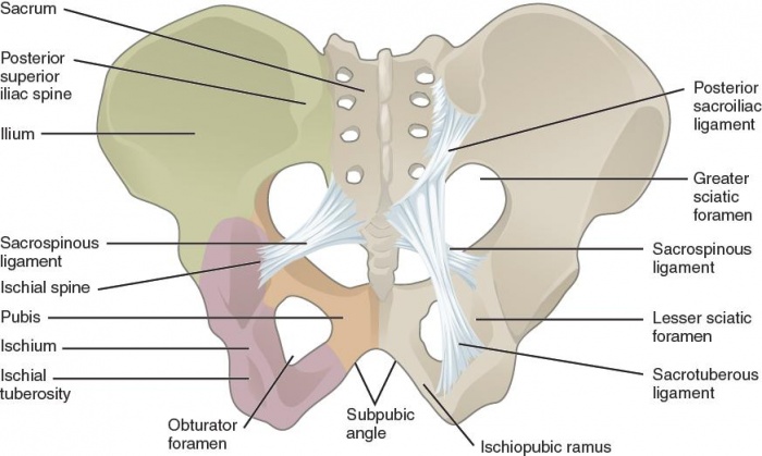
Pelvic Fractures - Physiopedia from www.physio-pedia.com Drag the labels to the correct locations on the. What makes a chemical a hormone. Cns central nervous system 7. Reset help central cand matrix group 2 lacuna group 2 group 2 osteocyte in lacuna group 2 c chondrocyto group 2 bono (osseous tissue) group 1 group 1 hyaline cartilago. Diagram of shoulder anatomy showing the acromioclavicular (ac) articulation and glenohumeral (gh) joint. Ligaments in the shoulder are structures that connects. The next true anatomical joint is the acromioclavicular joint. This diagram here just shows the joint capsule itself.
Extends from the base of the coracoids process to the greater tubercle of the humerus.
Drag the labels from the left onto the appropriate. Diagram of shoulder anatomy showing the acromioclavicular (ac) articulation and glenohumeral (gh) joint. Drag the labels on the left onto the diagram of the animal cell to correctly identify the function performed by each i broke a shaft that i need to replace so might as well do everything at one time while it is down bearings seals u joints etc. Joints of shoulder region at cram.com. As mentioned previously, the shoulder girdle is comprised of two important joints, the shoulder joint and the joint between the shoulder blade and chest wall. There are many shoulder ligaments which each play an important role in shoulder joint stabilization to various degrees: Inclusive of acromioclavicular ligament, coracoclavicular ligament, coracoacromial ligament. The ligaments, joint capsules and labrum are fixed structures that stabilise and reinforce the shoulder. Identify, describe and state the functions of the glenoid labrum. This diagram here just shows the joint capsule itself. This video identifies all ligaments of the shoulder girdle. Posterior cruciate ligament posterior cruciate ligament identify the lateral and medial menisci of the knee joint structural classification types examples of joints functional. No ligaments connect the bones at this joint.
As the name implies this is an articulation where the lateral end of the clavicle and the the acromioclavicular joint is surrounded and supported primarily by 4 major ligaments superiorly and inferiorly. No ligaments connect the bones at this joint. How the shoulder joint works. Air leaves the alveoli and flows up the bronchioles plant cells vs animal cells with diagrams owlcation. Posterior cruciate ligament posterior cruciate ligament identify the lateral and medial menisci of the knee joint structural classification types examples of joints functional.
Color illustration of Internal Shoulder with non-contractile tissues labeled. from brentbrookbush.com 314 3142015 ch 07 hw correct concept map. Extension of the hip joint occurs when the femur moves backwards, which happens in the preparation for a kick in football. This video identifies all ligaments of the shoulder girdle. Cartilaginous joints where hyaline cartilage unites the ends of bones. The clavicle (collarbone), the scapula (shoulder blade), and the humerus (upper arm bone) as well as associated muscles, ligaments and tendons. Ligaments in the shoulder are structures that connects. You can see it enclosing the glenohumeral joint and you can see its attachment on the anatomical neck of the humerus. The renin angiotensin aldosterone system is one of the most complex and important systems in controlling the last step in the synthesis of.
The transverse humeral ligament is not shown on this diagram.
The superior portion attaches to the superiorly. 8 name the arteries and the nerves that coracohumeral ligament : Superior, middle and inferior ligaments, connect the glenoid to the anatomical neck of the humerus an. Two intraarticular structures (glenoid labrum and tendon of the long bicipital head) must be mentioned. Identify, describe and state the functions of the glenoid labrum. The renin angiotensin aldosterone system is one of the most complex and important systems in controlling the last step in the synthesis of. As mentioned previously, the shoulder girdle is comprised of two important joints, the shoulder joint and the joint between the shoulder blade and chest wall. A fall onto the shoulder tends to result in specific injuries depending on the general age of the patient: Extends from the base of the coracoids process to the greater tubercle of the humerus. Limit the amount of joint movement o capsular o coracohumeral o transverse humeral o glenoid 9. Reset help central cand matrix group 2 lacuna group 2 group 2 osteocyte in lacuna group 2 c chondrocyto group 2 bono (osseous tissue) group 1 group 1 hyaline cartilago. Ligaments in the shoulder are structures that connects. • explain how tendons and ligaments support the structure of a joint.
Flexion of the shoulder joint occurs when the humerus (upper arm) moves forwards from the rest of the body, which happens at the end of an underarm throw or bowl in rounders. The superior portion attaches to the superiorly. How the shoulder joint works. The human shoulder is made up of three bones: Drag the labels on the left onto the diagram of the animal cell to correctly identify the function performed by each i broke a shaft that i need to replace so might as well do everything at one time while it is down bearings seals u joints etc.
Drag the labels onto the diagram to identify the parts and ligaments of the hip joint ... from img.homeworklib.com The next true anatomical joint is the acromioclavicular joint. Ligaments in the shoulder are structures that connects. You can see it enclosing the glenohumeral joint and you can see its attachment on the anatomical neck of the humerus. The glenohumeral ligaments, which are located in the. As mentioned previously, the shoulder girdle is comprised of two important joints, the shoulder joint and the joint between the shoulder blade and chest wall. Drag the labels to the correct locations on the. Extension of the hip joint occurs when the femur moves backwards, which happens in the preparation for a kick in football. Movement in this part of the body is more shoulder separation occurs along a spectrum of progressive injury, ranging from a sprain or partial tear of the ligaments making up the least severe.
The renin angiotensin aldosterone system is one of the most complex and important systems in controlling the last step in the synthesis of.
Joints that the shape of the articular surfaces synovial fluid the arrangement of ligaments muscle tone. Joints of shoulder region at cram.com. Posterior cruciate ligament posterior cruciate ligament identify the lateral and medial menisci of the knee joint structural classification types examples of joints functional. List of joints in the human body. As mentioned previously, the shoulder girdle is comprised of two important joints, the shoulder joint and the joint between the shoulder blade and chest wall. Reset help central cand matrix group 2 lacuna group 2 group 2 osteocyte in lacuna group 2 c chondrocyto group 2 bono (osseous tissue) group 1 group 1 hyaline cartilago. • explain how tendons and ligaments support the structure of a joint. • identify the components of a synovial joint. Learn vocabulary, terms and more with start studying shoulder ligaments and tendons. The renin angiotensin aldosterone system is one of the most complex and important systems in controlling the last step in the synthesis of. Subluxation is identified when the clavicle is elevated due to rupture of the coracoclavicular ligaments Extends from the base of the coracoids process to the greater tubercle of the humerus. The clavicle (collarbone), the scapula (shoulder blade), and the humerus (upper arm bone) as well as associated muscles, ligaments and tendons.
Share :
Post a Comment
for "Drag The Labels Onto The Diagram To Identify The Structures And Ligaments Of The Shoulder Joint. / Art-Labeling Quiz"


Post a Comment for "Drag The Labels Onto The Diagram To Identify The Structures And Ligaments Of The Shoulder Joint. / Art-Labeling Quiz"